


 Foundational characteristics of cancer include proliferation, angiogenesis, migration, evasion of apoptosis, and cellular immortality. Find key markers for these cellular processes and antibodies to detect them.
 The SUMOplot™ Analysis Program predicts and scores sumoylation sites in your protein. SUMOylation is a post-translational modification involved in various cellular processes, such as nuclear-cytosolic transport, transcriptional regulation, apoptosis, protein stability, response to stress, and progression through the cell cycle.
 The Autophagy Receptor Motif Plotter predicts and scores autophagy receptor binding sites in your protein. Identifying proteins connected to this pathway is critical to understanding the role of autophagy in physiological as well as pathological processes such as development, differentiation, neurodegenerative diseases, stress, infection, and cancer.
GATA3 Antibody (Center)
Purified Mouse Monoclonal Antibody (Mab)
- SPECIFICATION
- CITATIONS
- PROTOCOLS
- BACKGROUND

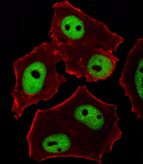
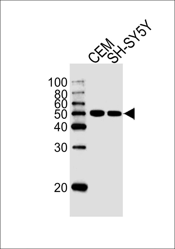
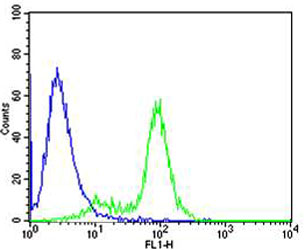
Application
| IF, FC, WB |
|---|---|
| Primary Accession | P23771 |
| Reactivity | Human |
| Host | Mouse |
| Clonality | Monoclonal |
| Calculated MW | H=48 KDa |
| Isotype | IgG2b,κ |
| Antigen Source | HUMAN |
| Gene ID | 2625 |
|---|---|
| Other Names | Trans-acting T-cell-specific transcription factor GATA-3, GATA-binding factor 3, GATA3 |
| Dilution | IF~~1:25 FC~~1:25 WB~~ 1:1000 |
| Target/Specificity | This GATA3 antibody is generated from a mouse immunized with a recombinant protein from human GATA3. |
| Format | Purified monoclonal antibody supplied in PBS with 0.09% (W/V) sodium azide. This antibody is purified through a protein G column, followed by dialysis against PBS. |
| Storage | Maintain refrigerated at 2-8°C for up to 2 weeks. For long term storage store at -20°C in small aliquots to prevent freeze-thaw cycles. |
| Precautions | GATA3 Antibody (Center) is for research use only and not for use in diagnostic or therapeutic procedures. |
| Name | GATA3 |
|---|---|
| Function | Transcriptional activator which binds to the enhancer of the T-cell receptor alpha and delta genes. Binds to the consensus sequence 5'-AGATAG-3'. Required for the T-helper 2 (Th2) differentiation process following immune and inflammatory responses. Positively regulates ASB2 expression (By similarity). Coordinates macrophage transcriptional activation and UCP2-dependent metabolic reprogramming in response to IL33. Upon tissue injury, acts downstream of IL33 signaling to drive differentiation of inflammation-resolving alternatively activated macrophages. |
| Cellular Location | Nucleus. |
| Tissue Location | T-cells and endothelial cells. |

Thousands of laboratories across the world have published research that depended on the performance of antibodies from Abcepta to advance their research. Check out links to articles that cite our products in major peer-reviewed journals, organized by research category.
info@abcepta.com, and receive a free "I Love Antibodies" mug.

Provided below are standard protocols that you may find useful for product applications.
Background
Transcriptional activator which binds to the enhancer of the T-cell receptor alpha and delta genes. Binds to the consensus sequence 5'-AGATAG-3'.
References
Joulin V.,et al.EMBO J. 10:1809-1816(1991).
Ho I.-C.,et al.EMBO J. 10:1187-1192(1991).
Ko L.J.,et al.Mol. Cell. Biol. 11:2778-2784(1991).
Marine J.,et al.Proc. Natl. Acad. Sci. U.S.A. 88:7284-7288(1991).
Deloukas P.,et al.Nature 429:375-381(2004).
If you have used an Abcepta product and would like to share how it has performed, please click on the "Submit Review" button and provide the requested information. Our staff will examine and post your review and contact you if needed.
If you have any additional inquiries please email technical services at tech@abcepta.com.
Ordering Information
Other Products
Shipping Information
